Smarter Scans, Better Care: How AI Is Revolutionizing Echocardiography
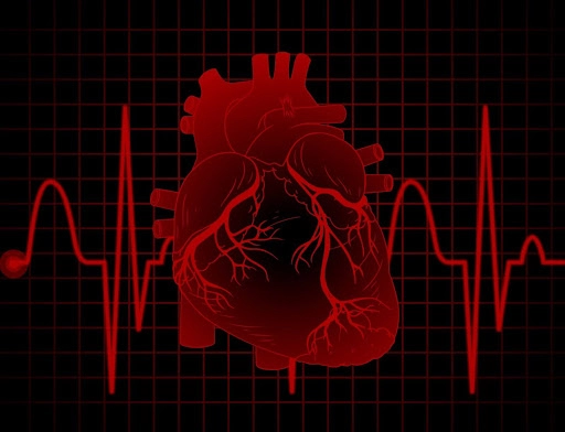
Cardiovascular diseases (CVD) claim over 20.5 million lives every year, according to the World Health Organization (WHO). Many of these deaths could be prevented by early diagnosis. Integrating Artificial Intelligence (AI) into echocardiography has made scans smarter and has transformed their predictive potential, helping to make detection faster and more accurate.
Why AI is changing the game
Traditional echocardiography was operator dependent, which means that the image quality and interpretation varied between clinicians, leading to inconsistent diagnoses. Decision-making required acquiring, analyzing, and reporting scans; also, experienced sonographers or cardiologists were not available in remote areas. All these translated into life-threatening situations.
Two factors that triggered the use of AI are
- Access to large datasets.
- Availability of easy-to-use AI tools.
Together, they allow computers to analyze heart scans, and AI, with its perspective, standardizes and accelerates decision-making.
AI’s expanding role in echocardiography and cardiology
New technologies are shaping echocardiography and automating processes using AI.
- Natural language processing (NLP), a tool in AI, reads and interprets unstructured text such as echo reports and clinical notes. It turns them into structured, usable data.
- Machine learning (ML) and deep learning (DL), combined with NLP, enable the measurement of heart functions, detection of valve problems, and prediction of outcomes.
AI analyzes these scans, providing quicker and more reliable care, quicker documentation, consistent diagnoses, less dependence on operator skill, and improved outcomes for patients.
AI in action: From data to diagnosis
The process begins with
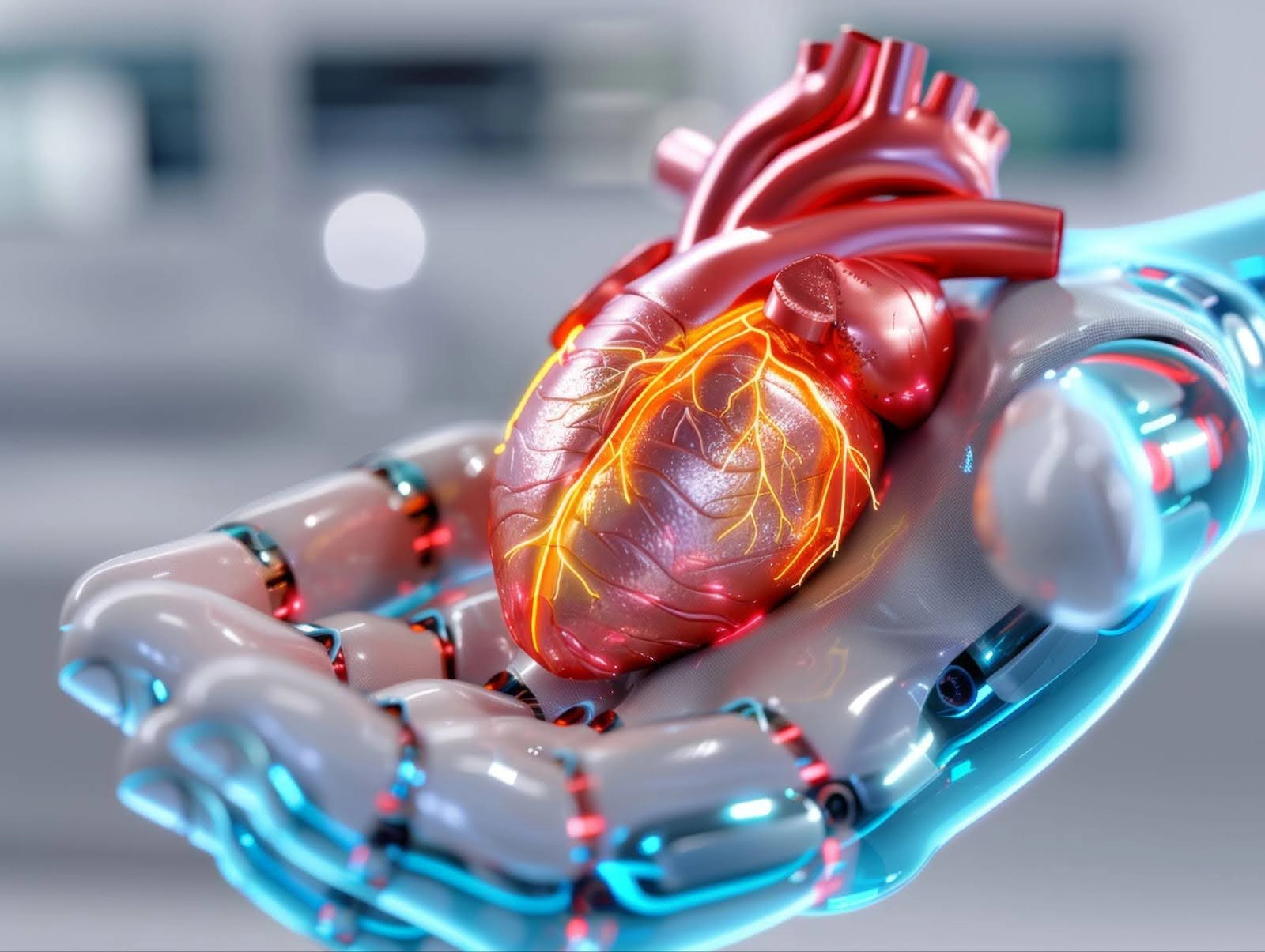
Smart data entry (data abstraction)
AI extracts clinical data and lab data from electronic medical records and prefills echocardiography systems. Prefilling reduces manual effort in manual entry, streamlines the workflow, and improves accuracy and efficiency.
Image acquisition
A shortage of trained sonographers means that AI-based echocardiographic tools help non-experts acquire high-quality cardiac images. Tools like UltraSight, Caption Health, and EchoGPS, integrated into echocardiographic systems, provide reliable images with minimal training.
Volumetric analysis
AI tools allow
- Fast, reproducible, and highly accurate calculations of the volumes of chambers of the heart and ejection fraction
- Myocardial strain and speckled tracking aid in analyzing the strain of the left ventricle, left atrium, and right ventricle; detecting tiny speckles in heart muscles facilitates early detection of dysfunction.
As technology advanced, the Echo CT (combination of 3D echo and AI-enhanced cardiac CT) was developed, while early tools could trace the heart’s inner walls and calculate how well the heart pumps blood. The new tool allowed doctors to view the heart from different angles, simulate surgeries, and focus on smaller areas like valves and chambers.
AI now rivals MRI in measuring right ventricular volume, making cardiac imaging more accessible and effective.
Wall motions
Analyzing the wall motion of the heart is rather difficult even for experienced doctors. It is so fast that the human eye cannot perceive the movements. AI-powered tools like EchoGo Pro analyze both resting and stress echocardiograms with accuracy comparable to that of expert humans. It will benefit the hospitals that lack top-level expertise but can still offer high-quality results.
Doppler and echocardiography from manual to automatic
Before the advancement of Doppler technology, doctors calculated the speed of blood manually. In contrast, modern ultrasound machines automatically calculate not only the speed of blood flow but also the pressure it creates. Advanced tools also measure mitral regurgitation when the blood leaks backwards. A color Doppler gives quick and accurate insights into heart function.
Smart assessment of the mitral valve
The mitral valve is a complex structure of the heart, and even small abnormalities can cause mitral regurgitation (MR), a condition where the blood leaks backward. Advanced AI tools measure the ring size, leaflet size, and shape automatically and expertly. The technology helps in improving both surgical and non-surgical repairs based on the specific needs of the patient's anatomy, utilizing 3D valve maps and measurements.
Smart aortic valve assessment
When the aortic valve becomes narrowed or undergoes stenosis, valve replacement is done. It requires measurements of the height of the coronary arteries and the size of nearby structures. 3D echocardiography with AI provides accurate measurements for these tools. These are trained on large datasets and can predict severe cases and survival outcomes with great accuracy.
AI sees what the human eye can’t
AI, using phenotyping (studying observable characteristics or phenotype results from the interaction between its genetic makeup and environment) and prognostication (predicting future events), spots features invisible to us. It can predict the age, gender, and risk of sudden death.
AI detects diseases that are hard to detect early
- Cardiac amyloidosis
- Coronary artery disease
- Hypertrophic cardiomyopathy
- Aortic stenosis
- Heart failure with preserved ejection fraction (HFpEF)
Understanding echo reports with AI
Echoinfer is a new tool that helps to mine echocardiographic reports and pull out details from the unstructured data. It aggregates several data points, including heart chamber size, valve problems, and blood flow measurements, and organizes them into a clear, usable format.
Using AI and natural language processing, researchers have built advanced data systems to turn echo reports into structured databases. This database helps to
- Integrate electronic health records
- Build large clinical registries
- Improve patient care
A heart scan revolution
Philips, Siemens, and TomTec lead innovation in the field of cardiology. AI will be the backbone of modern echocardiography, making it faster, easier, and more accurate. AI streamlines every step of the process: data entry, images, and accurate measurements. As AI evolves, it will revolutionize cardiovascular disease management and improve patient care.
While echocardiography is being done in many setups, not all patients have equal access to expert-level interpretation, especially in remote areas. AI is helping bridge that gap.
Inspired by:
Use of Artificial Intelligence Including Multimodal Systems to Improve the Management of Cardiovascular Disease
Biyanka Jaltotage, Juan Lu, Girish Dwivedi
Here to answer all your questions
An ECG measures the heart's electrical activity, capturing the timing and strength of the electrical signals that trigger heartbeats. This helps assess the heart's rhythm and detect potential abnormalities.
No, an ECG is not painful. The procedure involves placing electrodes on the skin, which may cause slight discomfort or irritation, but it does not include any electrical shock or invasive procedures.
Preparation is minimal. You may be asked to remove any jewelry or electronic devices and wear loose clothing to facilitate electrode placement. Avoiding heavy meals, caffeine, and smoking before the test may also be recommended.
An ECG is performed by placing small, sticky electrodes on the skin of the chest, arms, and legs. These electrodes are connected to a machine that records the heart's electrical activity and displays it as a series of waves on a graph.
A standard ECG test typically takes about 5–10 minutes. The preparation time for placing electrodes and recording is relatively quick and straightforward.
On an ECG, a PAC shows an early P wave with abnormal morphology followed by a narrow QRS complex. A PVC shows a wide, bizarre QRS complex (>120 ms) without a preceding P wave, because the signal originates in the ventricles and bypasses the normal conduction system.
In healthy individuals, both PACs and PVCs are generally benign. However, frequent PVCs (>10,000 per day) may lead to PVC-induced cardiomyopathy, and PACs can trigger atrial fibrillation in susceptible patients. A cardiologist should evaluate frequent or symptomatic premature beats.
ECG results are analyzed based on the pattern and timing of the heart's electrical signals. Abnormalities in these patterns can indicate issues such as arrhythmias, heart attacks, or other cardiac conditions, guiding further diagnosis and treatment.